
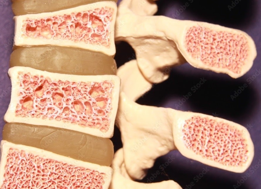
Bone mass typically peaks around age 35 and starts to decrease progressively thereafter. It has been estimated that 90% of women above the age of 75 are clinically diagnosed with osteoporosis.
The lifetime risk of a symptomatic osteoporotic fracture has been reported as 16% for females and 5% for males. Fractures of the vertebrae (spine), hips, and distal radius (wrist) are the more commonly encountered fractures related to osteoporosis, with osteoporotic spine fractures being the most commonly seen. These fractures may occur even without a history of a fall due to the fragility of the skeleton.
Patients with osteoporotic spine fractures, also known as osteoporotic compression fractures, may experience significant pain and reduced mobility.
Complications of such fractures include chronic pain, spinal deformity such as kyphosis (hunched back), spinal cord compression leading to paralysis or loss of bladder or bowel control, and breathing difficulties. Various studies have shown a 30% increase in mortality upon diagnosis of an osteoporotic spine fracture.
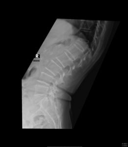
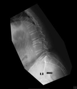
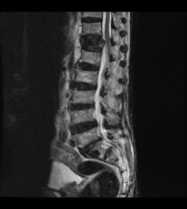
Investigations
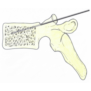
(i) Vertebroplasty
This procedure is a minimally invasive percutaneous (without surgical incision) technique in which the integrity and structure of a damaged or destroyed vertebral body is augmented through the use of bone cement. This procedure has gained widespread acceptance because of its efficacy and ability to be performed under sedation instead of general anaesthesia, thus allowing it to be performed as a day surgery procedure.
(ii) Kyphoplasty
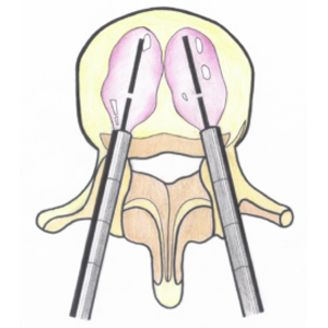
This procedure is an improved version of vertebroplasty, where an inflatable balloon is inserted into the fractured vertebra to create a void for cement filling (the balloon is removed prior to cement injection). This method further raises the safety level of the procedure, as the risk of cement leakage may be reduced significantly compared to vertebroplasty.
(iii) Stenting
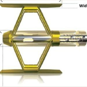
Stenting in the fractured vertebra reinforced by injection of bone cement around it is another modification of the vertebroplasty technique. The stent and the cement help maintain the integrity of the fractured vertebra.
Apart from managing the osteoporotic spinal fracture, it is important to treat the general osteoporosis condition as well. This can be achieved with various medications. Available options include oral weekly risedronate, six monthly injection of denosumab, daily injection of teriparatide or monthly injection of romosozumab.
Surgery
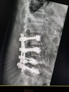
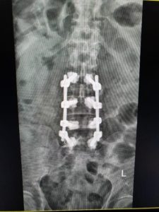
In severe cases of osteoporotic fractures causing neurological compromise, spine surgery may be required. This entails decompression of the nerves as well as stabilisation of the fractured vertebrae via titanium screws inserted above and below the fractured vertebra. Such screws may be reinforced with cement to strengthen the purchase of the screws into the vertebrae, helping to prevent implant failure.
When to Consult a Spine Specialist
Osteoporotic spine fractures may cause persistent back pain, spinal deformity, reduced mobility, and neurological symptoms. Early assessment by a spine specialist may help guide appropriate osteoporosis treatment and prevent worsening spinal collapse or nerve compression.

|
Dr Hee Hwan Tak
Orthopaedic Surgeon & Spine Specialist MBBS (Singapore) | FRCS (Edinburgh) | FRCS (Glasgow) | FAMS (Ortho) |
| View Dr Hee Hwan Tak’s Profile → |