
Introduction
Neck pain and back pain are among the most common musculoskeletal conditions seen by general practitioners and spine specialists.
A doctor will often have a good idea of the location and likely cause of a spinal condition after taking a careful clinical history and performing a directed physical examination. Imaging tests such as X-rays, MRI scans, and CT scans may then be ordered to confirm the findings before deciding on the most appropriate treatment plan.
X-rays
X-rays are often the first-line imaging test used in the evaluation of spinal conditions. They are widely available, cost-effective, and can be performed quickly and easily.
Abnormalities of spinal alignment are well demonstrated on X-rays, such as abnormal stepping between vertebral levels (spondylolisthesis) and abnormal spinal curvature (scoliosis) (Figure 1). X-rays are also commonly used to detect fractures following injury or trauma.
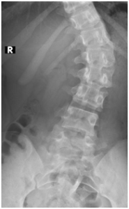
Figure 1. There is scoliosis of the spine which is curved to the left side. X-rays are useful for evaluating the severity of scoliosis, and repeat X-rays can be performed to monitor for progression. If there is an underlying congenital spinal deformity contributing to the scoliosis, it may also be visible on X-rays.
X-rays are useful for screening spinal degeneration, which may appear as bone spur formation in the vertebral bodies, enlargement (hypertrophy) of the facet joints, and disc space narrowing (Figure 2).
However, because X-rays only show the bony structures of the spine, soft tissue structures such as the intervertebral discs, spinal cord, spinal nerves, and surrounding muscles cannot be directly evaluated using X-rays alone.
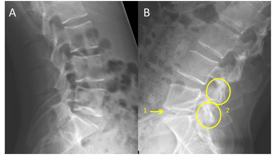
Figure 2. Picture A is an X-ray of a relatively normal lumbar spine in a young patient. Picture B shows degenerative changes including disc space narrowing (1) and facet joint hypertrophy (2). Disc space narrowing usually indicates underlying disc problem, although it does not reliably predict the severity. There may still be significant disc degeneration if the disc space is normal. MRI is still the most reliable test to evaluate discs.
MRI scan
MRI (Magnetic Resonance Imaging) is an advanced imaging scan that uses magnetic fields and radio waves without exposing patients to ionising radiation.
MRI allows detailed assessment of both bone and soft tissue structures of the spine and has played an increasingly important role in evaluating spinal conditions over the past few decades.
MRI is not required for all spinal conditions. However, if symptoms persist despite initial treatment or if there are signs suggesting a significant underlying spinal condition, a doctor may recommend an MRI scan.
MRI is highly useful for evaluating degeneration of the spinal discs, joints, nerves, and spinal cord. It can identify compression of the spinal cord or spinal nerves caused by bone spurs, disc prolapse, or other abnormalities (Figure 3).
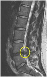
Figure 3. MRI shows that the L4-L5 disc is degenerated and protruding into the spinal canal, causing compression of the spinal nerves.
Occasionally, MRI may uncover less common but important conditions such as spinal infections, tumours, or spinal cord abnormalities that may not be visible on X-rays alone (Figure 4).
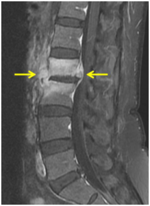
Figure 4. The patient experienced significant back pain over a period of time, which was eventually found to be caused by a spinal infection rather than disc degeneration.
CT Scan
CT (Computed Tomography) is an advanced imaging scan that uses X-rays to produce detailed cross-sectional images. It allows radiologists and spine surgeons to study the bony anatomy of the spine in great detail.
CT scans of the spine are less commonly performed than MRI scans, but there are situations where they are particularly useful. These include urgent evaluation of complex spinal fractures following major trauma (e.g. road traffic accidents), evaluation of the spine after surgery to assess bone healing and implant integrity, and guidance for certain spinal procedures (Figure 5).
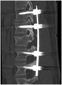
Figure 5. The presence of metal implants in the spine can significantly affect MRI image quality, whereas CT scans are less affected. Because CT scans provide excellent visualisation of bony structures, they are useful for assessing bone healing and detecting implant loosening or breakage.
CONTRIBUTED BY DR LEE CHIN HWEE, CONSULTANT RADIOLOGIST
When to Consult a Spine Specialist
Persistent neck pain, back pain, numbness, weakness, or symptoms affecting daily activities may require further evaluation. A spine specialist can determine whether imaging tests such as X-rays, MRI scans, or CT scans are necessary to diagnose the underlying spinal condition and guide appropriate treatment.

|
Dr Hee Hwan Tak
Orthopaedic Surgeon & Spine Specialist MBBS (Singapore) | FRCS (Edinburgh) | FRCS (Glasgow) | FAMS (Ortho) |
| View Dr Hee Hwan Tak’s Profile → |